Differential Media:
Overview of an Isolation Procedure for
Edwardsiella tarda
with an Analysis of "ET Agar"

Updated with clarification of medium preparation and the related
pH-based differential media theory on June 15 and 16, 2018.

YOU ARE HERE:
John L's Bacteriology Pages >
Selected General Topics >
Differential Media >
"ET Agar" for Isolation of
Edwardsiella tarda
x

  • This web page purposely falls short of becoming an official journal publication, as the basic information concerning the usefulness of the novel medium, "ET Agar" in my 1991 presentation at an ASM Poster Session has already been referenced in several publications from laboratories that have found the medium useful in their own isolation work. Those laboratories are where I consider the actual confirmation of the medium's value to be taking place. (One can do a web search for "Edwardsiella tarda" along with "ET Agar." As an example, check out this article.) So, the idea and application of the medium has "gotten around" as such, and what is presented here can simply be another one of my Differential Media pages for analyzing a selective isolation medium intended for certain enteric bacteria – in this case, Edwardsiella tarda.
    The reference one finds for my ASM Poster Session is as follows: Lindquist, J.A. (1991) Medium and procedure for the direct, selective isolation of Edwardsiella tarda from environmental water samples. Abstr. Annu. Meet. Am. Soc. Microbiol. C-303, p. 302.

  • At the end of this page are links to two appendices in PDF:
    • A table of the characteristics of the isolates obtained during the course of my 1988-93 project which initiated the use of ET Agar as a selective-differential isolation medium.
    • An attempt at a note meant for a bacteriological journal, written in 1991. It is put here in a journal format and can be useful for a number of relevant references.

  • A paper meant for official publication which describes the isolation and characterization of a possible new genus found among the water isolates in the aforementioned project has been in the hands of the CDC for some time. With the prospect of publishing that paper in mind, I stop short on this web page of referring to any specific scientific name or going far into detail, and the designation "G30 Biogroup" will suffice herein. Thirty one isolates were obtained from six Wisconsin lakes. The CDC's designation of this organism as CDC Enteric Group 121 and its 16S RNA description are referenced in the summary table of isolate characteristics (i.e., the first appendix) linked at the end of this web page.
    The ASM Poster Session where this investigation was presented is as follows: Lindquist, J. A. and J. J. Farmer III. (1999) Isolation and characterization of a new genus of Edwardsiella-like bacteria from Wisconsin lakes. Abstr. Annu. Meet. Am. Soc. Microbiol. R-1, p. 616.

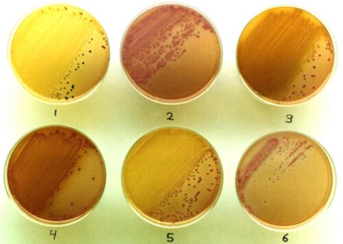
1. INTRODUCTION

Before its recognition as a taxonomic entity, Edwardsiella tarda was shown to cause disease in humans and a variety of warm and cold-blooded vertebrates. One would think that E. tarda isolates should be relatively easy to identify in a laboratory, as they are negative for fermentation of most sugars (including lactose, sucrose, mannitol and xylose), negative for phenylalanine deamination, and positive for lysine decarboxylation and hydrogen sulfide production. As this species is a member of the Family Enterobacteriaceae and also easy to cultivate, the colonies should come up well on a selective medium such as MacConkey Agar. Furthermore, this species is known to be resistant to the antibiotic colistin, a characteristic shared by few other enteric bacteria.

Keeping these characteristics in mind and remembering how the reactions from multiple substrates undergoing fermentation, deamination and decarboxylation can result in a net acidic or alkaline reaction for colonies on differential plating media (as demonstrated in the exercise here) or in such tubed media as TSI Agar and LIA, formulating a reasonably dedicated plating medium for the isolation of this organism should not prove to be difficult. If it works in theory as an appropriate selective-differential medium for E. tarda isolation, then it may very well work for culturing field samples. What follows shows how this came about.

x

2. DEVELOPMENT OF "ET AGAR" AND THE CHOICE OF "PRESUMPTIVE IDENTIFICATION" MEDIA

The following formula was developed and given the name "ET Agar." The H2S-indicator system of XLD Agar (Difco Laboratories, Detroit, MI) was incorporated. "Fine-tuning" was accomplished with some stock cultures and field samples. Indeed, concentrations and choices of the ultimate components of a selective-differential medium are adjusted as situations arise and results dictate. A small amount of additional agar was utilized for awhile (and may show up in some references), but this further solidification of the medium was determined unnecessary. Ultimate results involving six colistin-resistant enteric genera are shown farther below.

Ingredients
(Each is listed with its final concen-
tration
to be achieved per liter of
the completed medium.)
Reasons and expectations for use in detecting Edwardsiella tarda
MacConkey Agar Base (40 g/L)
Yeast Extract (1 g/L)
Glucose (2 g/L)
Colistin (5-10 mg/L)
x
  • Selection for gram-negative bacteria. These include the "enterics" which are expected to grow well in the presence of the selective agents in MacConkey Agar.
  • The pH indicator in the MacConkey base shows red (for net acidity) or white (for net alkalinity).
  • Basic nutrients. The peptone content of the MacConkey Agar base (containing amino acids which may be aerobically deaminated) is 2%. A bit of yeast extract is included as an additional nutrient source.
  • Glucose (added in a 0.2% concentration). There is just enough glucose for fermentation to occur that would tend to over-neutralize the alkaline effect of aerobic amino acid deamination. This concentration works to give Morganella colonies a net acidic reaction for better differentiation from E. tarda; see photo in table of results below for ET Agar where E. tarda has white, alkaline colonies and the rest have red, acidic colonies.
    (By comparison, the inability of 0.1% glucose in the TSIM – which, being based on TSI and KIA also contains 2% peptone – to counteract this deamination is shown below in the discussion of presumptive identification media.)
  • Selection for colistin-resistant bacteria. (Most enterics are sensitive to this antibiotic.)
Sucrose (5 g/L)
Mannitol (5 g/L)
Xylose (5 g/L)
L-lysine (10 g/L)
x
  • Typical E. tarda strains decarboxylate lysine and do not ferment these sugars. Fermentation of the aforementioned small amount of glucose alone would not produce enough acid to over-neutralize the alkaline effect of lysine decarboxylation.
    Result = alkaline (whitish) colony.
  • Enterics other than E. tarda would tend to ferment one or more of these sugars, generally resulting in acidic (red) colonies.
Sodium thiosulfate (6.8 g/L)
Ferric amm. citrate (0.8 g/L)
x
  • E. tarda produces H2S from thiosulfate which reacts with the iron.
    Result = black center of colony.

Preparation and Use.

As heating all of the ingredients together is not advised, the medium is meant to be prepared in two parts as follows for a total of one liter:

  • Mixture A:  The MacConkey Agar Base and yeast extract are mixed into 900 ml of distilled water and autoclaved for 10 minutes, followed by cooling in a 50°C water bath.
  • Mixture B:  The remaining ingredients (sugars, lysine, colistin and the H2S indicator system) are solubilized in 100 ml of distilled water which is then filter-sterilized and added to Mixture A (at 50°C), thus achieving the final concentrations noted in the formula above for all of the ingredients.

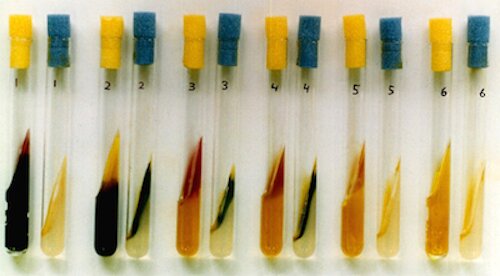
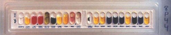
Usually, agar-containing media are initially steamed to melt the agar such that all the ingredients can be mixed thoroughly prior to autoclaving. However, I summarily decided from the outset to break tradition by not performing this pre-autoclave steaming of Mixture A, with the anticipation of doing a good mixing after the short, 10 minute autoclaving. From previous media-making experience, the mixing of the agar would be thorough enough, and overheating (however judged) should be avoided anyway. It was of great importance that Mixture A should attain plate-pouring temperature (about 50°C) before adding Mixture B, as a higher temperature might be deleterious to the sugars and other ingredients.

A Required Modification of the Preparation (once the project was under way).

As even some complete enteric plating media (SS Agar, for example) are not autoclaved but simply steamed to melt the agar, I was required to stop autoclaving Mixture A. This was after the project was well under way with its sampling, plating, isolating, testing, etc. Why was this so? With the advent of extreme automation in the Department's media facility, our old but reliable manual autoclave was summarily banished to the attic with other articles to eventually participate in the demolition/reconstruction project going on in that part of campus.

The unexpected happy result was that the medium, now prepared with simple steaming of Mixture A (prior to the addition of Mixture B), then began to support the additional growth of a new type of colony which was similar to that of E. tarda. More about this development is discussed appropriately in the "Isolations" section below (under "Exceptions").

Reactions Expected on ET Agar and Presumptive Identification Media for Selected Colistin-Resistant Enterics.

An overview of the results from the testing of representatives of six genera of colistin-resistant members of the family Enterobacteriaceae follows, but it is preceded by the following table of expected reactions on ET Agar and from substrates which could conceivably be incorporated into suitable "presumptive identification" media. Not all non-Edwardsiella species are specifically considered, and some strains of Proteus may conceivably produce black colonies resembling those of E. tarda. So, one would depend on the subsequent use of suitable screening media, and the choice was made of Phenylalanine Agar and Triple Sugar Iron Agar. The latter medium is modified with the addition of 1% mannitol which strains of E. tarda would not be expected to ferment; thus the designation "TSIM."

  E. tarda Proteus Morganella Providencia Serratia Cedecea
E
T

A
G
A
R

r
x
n
s
Glucose Fermentation + + + + + +
Sucrose, Mannitol
and/or Xylose Ferm.
+ – or + + +
Lysine Decarboxylation
(an alkaline reaction)
+ +
Net pH of Colony
(white=alk; red=acidic)
white red red red red red
H2S from Thiosulfate + +
H2S seen in Colony + –*
T
S
I
M

&

Φ
A
L
A
Glucose Fermentation + + + + + +
Lactose, Sucrose
and/or Mannitol Ferm.
+ or – – or + + +
Net pH of TSIM Slant**
(red=alk; yellow=acidic)
red yellow or red red red or yellow yellow yellow
H2S from Thiosulfate + +
H2S seen in TSIM Butt + +
Phenylalanine Deam.
(+ = green color with FeCl3)
+ + +

  * = The pH is too low (especially from xylose fermentation) for the black FeS precipitate to form.
** = The slant region would only show an acidic reaction diffusing through the medium from fermentation of lactose, sucrose and/or mannitol. If the glucose (in 0.1% concentration) were the only sugar fermented, there would not be enough acid expected to overneutralize the aerobic, alkaline deamination reaction; this is carrying on the same feature shown in the traditional media KIA and TSI on which TSIM is based.

Reactions Observed on ET Agar and Presumptive Identification Media for Selected Colistin-Resistant Enterics.

x

The cultures are numbered as on the table above:
1 - Edwardsiella tarda
2 - Proteus sp.
3 - Morganella sp.
4 - Providencia sp.
5 - Serratia sp.
6 - Cedecea sp.

On the ET Agar plates, note the distinguishing black-centered colonies and alkaline appearance (no red color) for E. tarda. What little acid is produced from the only sugar fermented by E. tarda (glucose) is easily overneutralized by the lysine decarboxylation reaction.

In the TSIM tube (yellow plug), a net alkaline (red) reaction (from aerobic deamination of amino acids) appears in the slant region for those which do not produce enough acid from fermentation to diffuse into this reagion to produce a net acidic (yellow) reaction. For E. tarda, a bit of acid is produced from glucose fermentation (with none from the other three sugars) and it is well over-neutralized by the alkaline reaction produced by amino acid deamination in the aerobic region.

Assisting in the differentiation of these organisms (especially if the pH reactions are not convincing enough) is the Phenylalanine Agar (blue plug). However, typical E. tarda isolates would be expected to hold true to what is seen here for any of its reactions on this table, and pursuing the identity of other colistin-resistant organisms is not the problem here.

x

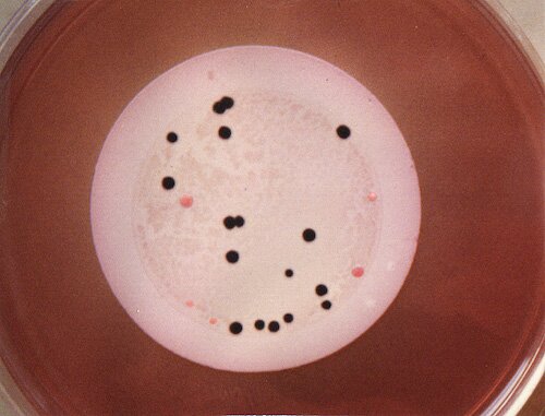
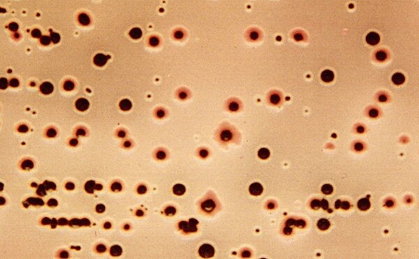
3. ISOLATIONS

In 1988, I began an analysis of Midwestern stream and lake samples for Edwardsiella tarda, passing various volumes of freshly-taken and refrigerated samples (usually 1, 4, 10 and 25 ml from each sample site) through 0.45 µm membrane filters (with the use of a suction flask) which were each placed upon a plate of ET Agar. One-day incubation at 37°C was generally sufficient for good colony formation. A typical plate showing good isolation of presumptive E. tarda colonies is shown below. With exceptions noted below, (1) colonies remained black (with also a whitish, alkaline rim) after subsequent streaking on a fresh plate of ET Agar (without colistin), and (2) the presumptive identification tests for the black colonies (detailed above) turned out positive for E. tarda.

x

Exceptions:

  • When isolates of the "new type of colony" mentioned above (i.e., that which arose after the preparation adjustment and was similar to E. tarda) were characterized, they were found to possess uniform, non-E. tarda-like characteristics in the media for presumptive and further identification. They were collectively established as the "G30 Biogroup," so-named after the first isolate. Distinguishing characteristics which differentiate that group from E. tarda are included in the summary table linked at the end of this web page. As noted in the beginning of this page, extensive details about the testing and naming of this unique group of bacteria go beyond the subject of E. tarda isolation and are most appropriately dealt with in a "for-real" publication which should happen in due course.

  • Another significant exception came from the occasional presence of generally smaller, black colonies that were ultimately identified as Shewanella putrefaciens which metabolizes primarily by means of respiration (aerobic and anerobic) and occasionally fermentation.

  • Very small, black colonies appeared from some samples which were ultimately identified as E. tarda but reacting very weakly in the test for hydrogen sulfide in TSIM. Upon further testing, I considered these isolates to be a new, tentative biogroup which I designated the "CL Biogroup" as I had initially noted them from a Clear Lake, Iowa sample. (I mentioned at my 1991 ASM poster session that they didn't react with the usual bacteriophage as those which had the typical reactions and regular-sized colonies. Since then, I lost the phage due to my having forgotten to include certain ions in the suspending solution. Phage work may eventually start again.)

  • On filter plates where the sample had been taken from the shore of a Lake Superior bay (near Ashland, WI) which contained a lot of dead fish and seagull feces, I noted some exceptionally reddish colonies which, when streaked on ET Agar, formed black colonies with a red (acidic) rim. This turned out to be identified as an E. tarda "Biogroup 1" isolate and confirmed so by the CDC. The biogroup is officially recognized as such in Bergey's Manual and other publications for its significant fermentation of sucrose and mannitol. However, when Bergey's Manual and others state that the H2S reaction is negative for Biogroup 1, they do not make clear that their test medium is TSI Agar. It is indeed negative in TSI Agar, but not in the API-20E test or on Kligler Iron Agar where there is no sucrose to be fermented to produce the excess acid to negate the H2S reaction. In the API-20E test (shown below), the Biogroup 1 isolate was identified as Edwardsiella hoshinae, probably because Biogroup 1 was not yet in the database. The isolate could have been ignored from the outset but for a certain curiosity of the experimenter regarding its actual identification – which certainly made it a "keeper."

x

A demonstration plate on ET Agar was prepared from a mixed suspension of cells from three isolates, each representing a biogroup of E. tarda. The normal type of colony is black with a whitish rim. The CL Biogroup is seen as the tiny dark colonies, and Biogroup 1 is recognized by the red (acidic) rim due to its fermentation of sucrose and mannitol.

x

APPENDIX 1.  SUMMARY TABLE OF THE "BIOGROUPS" AND THEIR DIFFERENTIATING REACTIONS

A table showing the biochemical reactions of the several biogroups is on a convenient PDF file which can be accessed here. Included on the table is a column detailing the general reactions of the aforementioned "G30 Biogroup" which I have often characterized as "trying to pass itself off as Edwardsiella tarda" on the original filter plates. However, it is certainly different!

APPENDIX 2.  A NOTE FOR A BACTERIOLOGICAL JOURNAL WRITTEN IN 1991

"A Medium for the Isolation of Edwardsiella tarda" was drafted as a possible note for publication but was set aside, mainly due to the volume of work at my job as a lab instructor at U.W.-Madison. It is probably of value for its references to background material on Edwardsiella tarda. The PDF copy can be found here.


Return to the main page of
the Differential Media site.


Page slightly modified (for clarity) on 7/14/18 at 5:00 PM, CDT.
John Lindquist:  home page and complete site outline

E-mail me at  jlindquist 001 @ gmail.com
Be sure to remove spaces (added to discourage spambots).
Department of Bacteriology, U.W.-Madison